Biopsy

Biopsy is usually performed for the purpose of genetic testing that can greatly improve the chance of achieving a successful pregnancy.

During the last decade, the main objective of assisted reproduction technologies (ART) evolved from achieving a pregnancy, to achieve a healthy live newborn.

Between 40% and 75% of human embryos may contain an abnormal number of chromosomes, depending on the woman’s age. Most chromosomally abnormal embryos will not implant and if they start to implant are most likely to miscarry.
One of the main goal of reproductive medicine laboratories is to maximize the number of live births per stimulated cycle helping patients to reduce the costs, time and efforts to achieve a pregnancy.
To achieve this goal, efficient embryo selection strategies must be applyed to identify those that are reproductively competent within a cohort obtained during an IVF cycle.
There is evidence that blastocyst culture combined with genetic testing and transfer of vitrified and warmed euploid embryos is an approach that could increase efficiency.
Some researchers suggest that pre-implantation genetic screening could be used as a clinical tool to improve implantation and live birth rates and, in turn, decrease multiple pregnancies and the risk of miscarriage.

Preimplantation genetic testing (PGT) aids selection of embryos that are euploid or that are lacking any particular genetic disorder for embryo transfer and further improve IVF outcomes (1). Biopsy is performed by removal of 1 or 2 blastomeres on day 3 embryos or 5-10 trophectoderm cells on the blastocyst stage (day 5-7).

(1)Rodriguez-Purata J. et al. Reproductive outcome is optimized by genomic embryo screening, vitrification, and subsequent transfer into a prepared synchronous endometrium. J Assist Reprod Genet. 2016 Mar; 33(3): 401-412.

Studies have shown that implantation and clinical pregnancy rates are higher in deferred transfers, indicating that the reproductive outcome is optimized with embryos with genetic screening, vitrification, and subsequent transfer into a prepared synchronous endometrium (1).
Trophectoderm biopsy is the most common method of embryo biopsy for pre-implantation genetic testing (PGT). Reducing laser exposure to the embryo and procedure time are the main goals for any practitioner when biopsying. Because of this, different approaches to the technique have been developed. Once the embryo is held, the biopsy micropipette will pull 5-10 trophectoderm cells into the pipette and laser shots fired at cell junctions. Cells are pulled apart mechanically while keeping pulling the cells with the biopsy micropipette.
For hatched blastocysts, flicking the embryo has been proposed as an alternative. After performing laser shots, both micropipettes are overlapped and tension on the biopsy micropipette, which is holding the embryo from the cells are aimed to biopsy, is created by moving the holding pipette. A quick movement, a flick, helps to disggregate the biopsy from the embryo.
In the case of PGT programs it is important to carry out a safe biopsy technique, but it is essential to implement a successful vitrification procedure to allow sufficient time to obtain the genetic result that will ensure the future viability of the embryo.


Although the prevalence of failed genetic tests is low, this will imply to re-biopsy the non-diagnosed blastocyst. Repeating the test will involve a second round of biopsy and a second round of vitrification. On the other hand, there are patients who can also request genetic tests on non-biopsied embryos that are already cryopreserved to allow a transfer of an euploid embryo. In this case, even though a single biopsy is performed, this will require two cryopreservation procedures.

Scientific evidence has demostrated that warming previously vitrified embryos followed by biopsy and re-freezing maintains embryo viability and clinical pregnancy rates (2). Furthermore, given that blastocyst re-biopsy produces an euploid result in a high percentage of cases and favorable pregnancy outcomes, this strategy deserves consideration for all patients who want to know the ploidy status of their entire cohort of embryos, and particularly for those patients who do not have other embryos available for transfer (3).

(1)Rodriguez-Purata J. et al. Reproductive outcome is optimized by genomic embryo screening, vitrification, and subsequent transfer into a prepared synchronous endometrium. J Assist Reprod Genet. 2016 Mar; 33(3): 401-412.

(2) Wilding M. et al. Thaw, biopsy and refreeze strategy for PGT-A on previously cryopreserved embryos. Facts Views Vis Obgyn. 2019, 11 (3): 223-227

(3) Neal S.A. et al. When next-generation sequencing-based preimplantation genetic testing for aneuploidy (PGT-A) yields an inconclusive report: diagnostic results and clinical outcomes after re biopsy. JARG. 2019, 36:2103-2109.

Biopsy dish set up

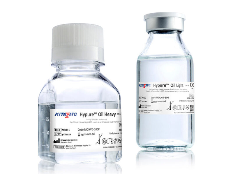
Biopsy dish set up is set up with Gamete Buffer droplets covered with Hypure® Oil. The embryo is firmly stabilized with the Holding Pipette while using the Biopsy Pipette to accurately remove biopsied cells. 

There are different strategies for opening the zona pellucida:  

– Mechanically with PZD Pipettes. 

– Chemically with an acid solution. 

– Laser shots.  

Kitazato Biopsy Pipettes

Are specially designed with a flat and polished tip to protect cell membrane during removal of blastomeres or trophectoderm fragments and minimizing harm to the biopsied embryo.

Kitazato PZD Pipettes

Are specially designed with a very sharp and thin tip to allow a fine cut and ease penetration of the zona pellucida without damaging the embryo.

The IVF Lifecycle

Kitazato offers a well curated selection of quality products that maximize success at every step of the IVF Lifecycle. Learn more about the products involved in each IVF procedure.

View all
Oocyte Retrieval
More info
Andrology
More info
Culture
More info
Vitrification
More info
Embryo Transfer
More info
Oocyte Retrieval
More info
Andrology
More info
Culture
More info
Vitrification
More info
Embryo Transfer
More info