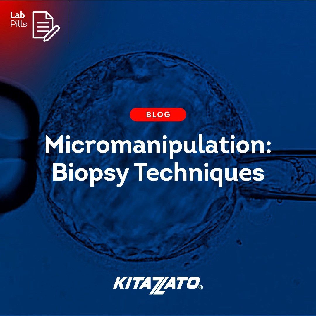
From laboratories to clinics, the main purpose of every professional involved in an IVF treatment is to deliver to our patients a healthy baby at home. To do so, many techniques and advancements have arisen over the past years. One of the revolutionary and revealing techniques in the IVF world is with no doubt, the trophectoderm biopsy.
How can biopsy techniques lead to successful outcomes?
If we look carefully to published data, we can affirm that at 38 years of age over 50% of the embryos are chromosomally abnormal. To increase the chances of an embryo transfer to be successful, the clinic can offer to the patients a PGT, always individually reviewing the case.
In the last decade, and thanks to the excellent results with vitrification among other advances, efficient and successful PGT programs have been established around the world. Many studies prove today that vitrification survival rate of PGT embryos are comparable to non PGT embryos.
So, with strong PGT and vitrification programs, outcomes in clinical pregnancy and live birth rates excel, as evidenced in the following chart.

There are several approaches to PGT, however, the trophectoderm biopsy is the most widely used worldwide for the simple reason that it gives the most accurate results. Blastocyst biopsy is characterized by:
- Analysis of multiple cells.
- It does not damage the internal cell mass (ICM) from which the fetus derives.
- Detects aneuploidies originated by both parents, structural anomalies of maternal and paternal origin and gene mutations.
A specialized procedure for any case
There are different effective approaches to trophectoderm biopsy depending on the different characteristics of blastocysts.
- If we have a fully expanded blastocyst, we can artificially shrink it with a laser shot on the junction between trophectoderm cells, then we will open a hole in the pellucid zone (ZP) to introduce the biopsy pipette and gently aspirate some cells.
Once the desired number of cells are inside the pipette, we will apply two or three laser shots and, by mechanical blunt dissection, will separate these cells from the rest by flicking the holding and the biopsy pipette with a quick and precise movement.

💡 Tip & Trick: In case the artificial shrinkage doesn’t work, we can shoot the laser in the zona pellucida and then blow some media inside the blastocoele, forcing this shrinkage, to then proceed with the biopsy and the mechanical blunt dissection.
- In case of hatching blastocyst, peanut shaped, we will shoot with the laser on the trophectoderm on the part that is outside the ZP. The embryo will shrink and then, we can perform the biopsy the same way as an expanded blastocyst, aspirating the cells through the remaining ZP as seen in the pictures.

💡 Tip & Trick: If at warming, the embryo doesn’t detach from the Cryotop, at the Thawing Solution step use another 35 mm dish full of Dilution Solution, to transfer the whole Cryotop in DS instead of trying to detach the embryo otherwise.
- For 8-shape hatching blastocysts, the embryo is secured by the holding pipette, and we can perform the biopsy directly in the cells outside of the ZP like in a fully expanded blastocyst. If the ICM is within the cells outside of the ZP we will perform the peanut shape blastocyst approach.
💡 Tip & Trick: It is also significant to highlight the importance of leaving extra Vitrification Solution media when vitrifying PGT embryos, specially hatched ones that had no protection from the ZP. If at warming, the embryo doesn’t detach from the Cryotop, at the Thawing Solution step use another 35 mm dish full of Dilution Solution, to transfer the whole Cryotop in DS instead of trying to detach the embryo otherwise.
Experience is essential, but high-quality tools are critical
As we have seen in previous blog posts, experience, and the precise execution of techniques by professionals in laboratories is crucial to ensure accurate and reliable results in the scientific field.
👀 Blog Post > Crafting excellence: accurated manipulation for art and pgt
The expertise acquired over time contributes to the efficiency and accuracy of procedures, ensuring the validity of the data obtained. However, it is essential to recognize that excellence in performance is not solely dependent on individual skill.
Excellent procedures as The Cryotop Method®, the use of high-quality tools and ongoing training are equally indispensable factors. Advanced and precise tools such Kitazato micropipettes, enhance the quality of results, while continuous training allows individuals to stay updated on the latest methodologies and technological advances.
Commitment with science
For this reason, Kitazato is committed to continuing to provide the best combination for its customers through virtual and face-to-face training worldwide, providing the necessary foundation for achieving high standards of excellence in laboratories and contributing to the advancement and reliability of scientific research.
As part of this commitment to excellence, Kitazato recognises those clinics and laboratories with outstanding results in their vitrification processes, thanks to the Best Practices Recognition Program. More than 50 clinics and laboratories are already part of this exclusive community.
👉 Find out more by clicking on the following link: IVF Lifecycle: Vitrification
